
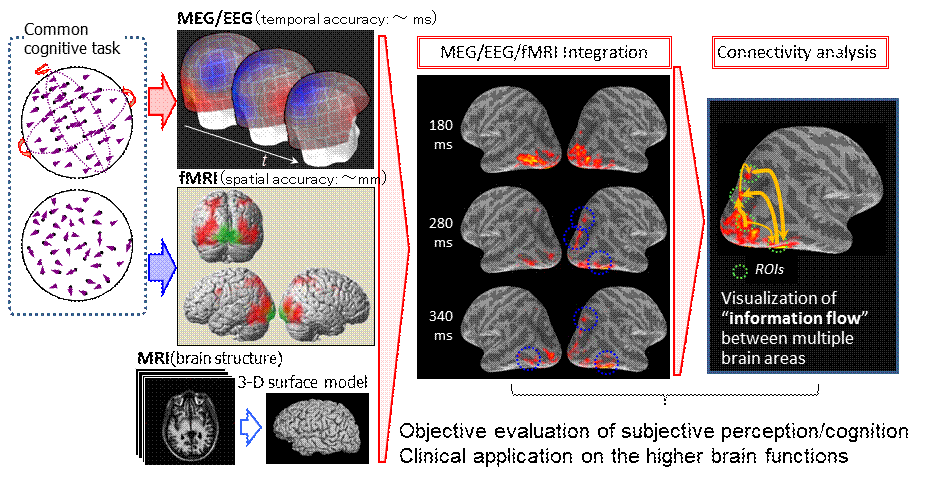
Multimodal Neuroimaging Technique and Connectivity Analysis
Each non-invasive neuroimaging modality has its own inherent limitations resulting from temporal and spacial inaccuracies due to the nature of information that can be measured. While functional magnetic resonance imaging (fMRI) techniques provide excellent spatial resolution up to sub-millimeter resolution, its temporal resolution is limited by the hemodynamic time constant. Conversely, magnetoencephalography (MEG) and electroencephalography (EEG), which measure temporal changes in neural current directly, have temporal resolution of approximately a few milliseconds. However, their spatial accuracy is limited by the non-unique nature of the problem of estimating the spatial distribution of neural currents from the measurement of voltage (EEG) or magnetic field (MEG) distributions from outside of the brain (inverse problem).We developed a technique to incorporate structural and functional MRI data into the MEG/EEG inverse procedure (noninvasive multimodal neuroimaging). The current results demonstrate that the noninvasive multimodal neuroimaging, in which structural and functional MRI data are integrated into the MEG/EEG inverse problem, is capable of visualizing neural dynamics of the human brain with spatial accuracy comparable to fMRI without compromising the excellent temporal accuracy of MEG/EEG.
Using the MRI/fMRI incorporated MEG/EEG inverse procedure, the time-course of neural activity can be extracted from multiple brain regions-of-interest (ROIs) with millisecond precision. These data could be used to infer neural interactions between various brain regions (connectivity analysis), which are considered to be crucial neural mechanisms underlying higher order brain functions specific to humans.
- Iwaki S., Multimodal neuroimaging to visualize human visual processing, Biomedical Engineering and Cognitive Neuroscience for Healthcare, IGI Global Press, pp. 274-282, 2012.
- Iwaki S., Sutani K., Visualization of the sensitivity of the MEG sensor array based on the 3-D modeling of cortical surface and volume conductor, J. Appl. Phys., 107: 09B317, 2010.
- Iwaki S., Bonmassar G., Belliveau J.W., Spatiotemporal imaging of the brain activities during 3-D structure perception from motion, Frontiers in Human Brain Topography, Elsevier, pp. 209-212, 2004.
Studies on Human Visual Functions
Neural Dynamics of 3-D Structure Perception from Motion
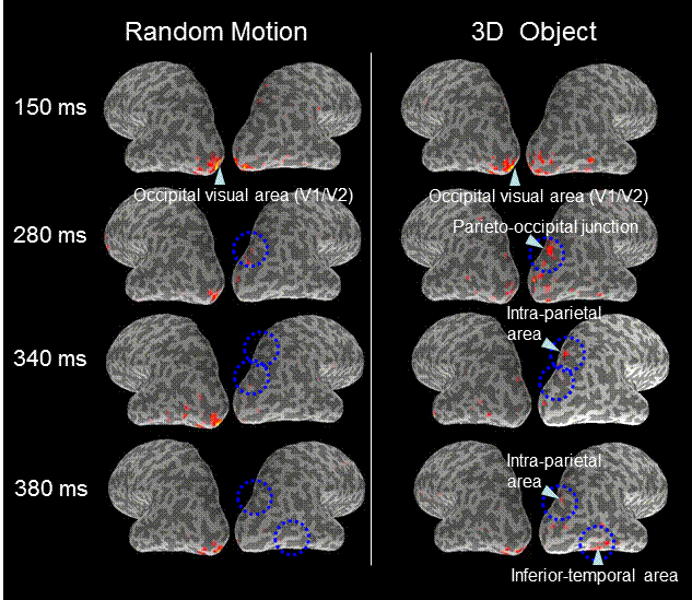
 During naturally occurring three-dimensional (3-D) object perception, shapes are often recognized as they move. Recent neuroimaging studies suggest involvement of both dorsal (i.e. the parieto-occipital (PO) and the intraparietal (IP) regions) and ventral (i.e. the posterior inferior temporal (IT) region) visual systems, as well as the motion processing area MT, in the perception of 3-D object from motion. However, the neural dynamics underlying the reconstruction of a 3-D object from 2-D optic flow is not known.
We combined neuromagnetic (MEG) and the hemodynamic (fMRI) measures to detect the dynamic brain responses underlying 3-D structure perception from 2-D motion in humans. This spatiotemporally accurate multimodal neuroimaging technique revealed the exact neural dynamics in the dorsal and ventral visual pathways.
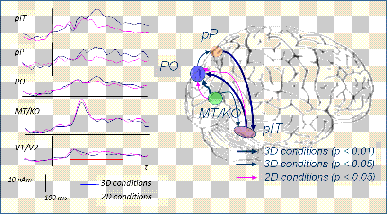
The interactions inferred by the connectivity analysis showed that there were significant directional influences from MT to PO, IP to pIT, and pIT to PO only during the perception of 3-D object structure with highly coherent random-dot motion conditions but not during the random motion perception. These results suggest that the perception of a rotating 3-D object from 2-D random-dot motion includes integration of global motion and object recognition, that are accomplished by the cooperative engagement of the ventral and the dorsal visual information processing streams.
- Iwaki S., Bonmassar G., Belliveau J.W., Dynamic cortical activity during the perception of three-dimensional object shape from two-dimensional random-dot motion, J. Integr. Neurosci., 12: 355-367, 2013
- Iwaki S., Belliveau J.W., Neural interactions between dorsal and ventral visual subsystems while perceiving 3-D structure from 2-D motion, Advances in Biomagnetism, Springer, pp. 286-289, 2010.
- Iwaki S., Bonmassar G., Belliveau J.W., Event-related changes in the spontaneous brain activity during 3D perception from random-dot motion, Complex Medical Engineering, pp.499-510, 2007.
Brain Activities during Mental Image Processing
COMING SOON ...- Nishimura K., Aoki T., Inagawa M., Tobinaga Y., Iwaki S., Mental rotation ability and spontaneous brain activity: a magnetoencephalography study, Neuroreport, 31: 999-1005, 2020. DOI 10.1097/WNR.0000000000001511
- Iwaki S., Sutani, K., Inagawa M., Tobinaga Y., Nishimura K., Performance-dependent changes in gamma-band brain activity during mental image processing, Soc. Neurosci. Abstr., #96.15, 2012.
- Iwaki S., Tonoike M., Ueno S., Visualization of the brain activity during mental rotation processing using MUSIC-weighted lead-field synthetic filtering, IEICE Trans. Inf. & Syst., 85-D: 175-183, 2002.
- Iwaki S., Ueno S., Imada T., Tonoike M., Dynamic cortical activation in mental image processing revealed by biomagnetic measurement, NeuroReport, 10: 1793-1797, 1999.
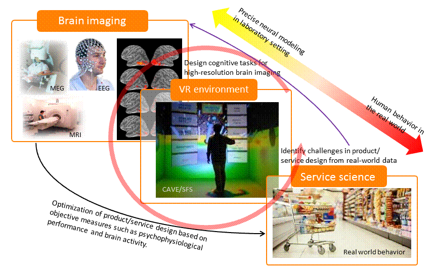
Development of "Neuro-Aided Design Framework" to Support Optimal Product/Service Design
Recent development of the behavioral economics have shown that our daily behavior as a consumer is not always rational and is influenced by subtle changes in the environment or in our own emotion which is sometimes below our conscious perception. In such situations, application of functional brain imaging techniques to optimize product and/or service designs, often referred to as "Neuroeconomics/Neuromarketing", has become a topic of broad attention.Although neuroeconomics have shown promising results to elucidate the neural mechanisms underlying our preference, decision making, and behavior, etc., it is still difficult to construct a model to predict our daily bahavior based on the neuroimaging data acquired non-invasively. One of the reason is that there is a significant gap between the laboratory experimental setting, typically in the MR scanner or EEG/MEG shielded room, and the real world environment where the service is actually provided. We are now trying to solve this issue by integrating brain imaging techniques with immersive virtual environment and real-world measurement of human behavior.
- Takeda Y., Okuma T., Kimura M., Kurata T., Takenaka T. Iwaki S., Electrophysiological measurement of interest during walking in a simulated environment, Int. J. Psychophysiol., 93: 363-370, 2014.
Mental Fatigue Measurement on the Personal Devices (Flicker Health Management System)
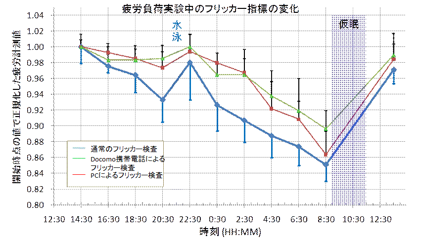
 Frequency domain flicker-perception threshold (FD-FPT) or critical flicker frequency (CFF) is defined as a frequency at which intermittency of light disappears into a steady state for a human observer. CFF is known to decrease as mental fatigue increases. Although the CFF has decades of history as a robust index of mental fatigue (flicker-test) in laboratory settings, it has not been widely used by the general public. This is primarily because it requires a dedicated device and measurement by a trained professional. Additionally, arbitrariness and subjectivity in subject responses in determining the ordinary flicker-test presents a significant problem for robust measurements of mental fatigue in everyday situations where there is no supervision for users on how to respond to the ongoing flicker stimuli.
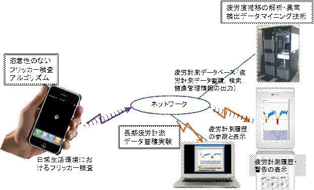
We developed (a) a new method to control a subjective flickering sensation by changing the contrast of the visual stimuli instead of changing temporal frequency, and (b) a new paradigm, in which forced-choice task performance was used to determine FPT to eliminate arbitrariness of subject responses. The proposed techniques were implemented as application software, which enables persons to measure mental fatigue quantitatively using consumer mobile devices such as smart phones and PCs (Flicker Health Management (FHM) System).
Efforts to bring these technologies to the market are being made at Flicker Health Management Corp, the AIST Start-ups based on AIST Technology Transfer.
- Iwaki S., Harada N., Mental fatigue measurement based on the changes in flicker perception threshold using consumer mobile devices, Adv. Biomed. Eng., 2, 137-142, 2013.
- Shichiri M. Harada N., Ishida N., Komaba L.K., Iwaki S., Hagihara Y., Niki E., Yoshida Y., Oxidative stress is involved in fatigue induced by overnight deskwork as assessed by increase in plasma tocopherylhydroqinone and hydroxycholesterol, Biol. Psychol., 94: 527-533, 2013.
- Iwaki S., Recent developments of non-invasive neuroimaging techniques and their possible application to quantitative evaluation of fatigue and stress, Stress Science Research, 28: 4-7, 2013. (In Japanese)
- Iwaki S., Harada N., Mental fatigue evaluation in the daily life based on the changes in flicker perception threshold, Function & Materials, 33(2): 4-8, 2013. (In Japanese)
Studies on Human Visuo-Motor Functions and Non-verbal Communication
Interactive Operatability of Human-Computer Interface
COMING SOON ...- Kashiwagi M., Iwaki S., Narumi Y., Tamai H., Suzuki S., Parietal dysfunction in developmental coordination disorder children: An fMRI study, Neuroreport, 20: 1319-1324, 2009.