Principle
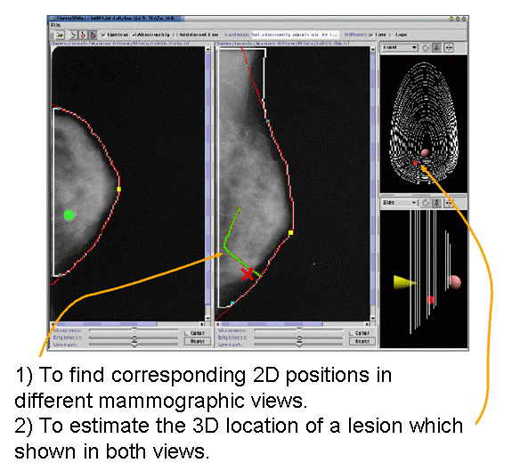
This figure shows an overview of our method [1)] , which enables us to calculate the the system. First, suppose that a point in one image is pointed at by a radiologist. The method calculates the epipolar curve, that is the locus of possible corresponding positions of the point in the other image, by simulating the five steps of the following process, A: back projection → B:uncompression → C: rotation → D: compression → E:projection, as shown by the solid arrows. Next, the corresponding position is searched for along the epipolar curve. Once the correspondence is found along the curve, the corresponding 3D position in the uncompressed breast can be determined by retracing the movement of the point during the simulation, as shown by the dashed arrows.

Results
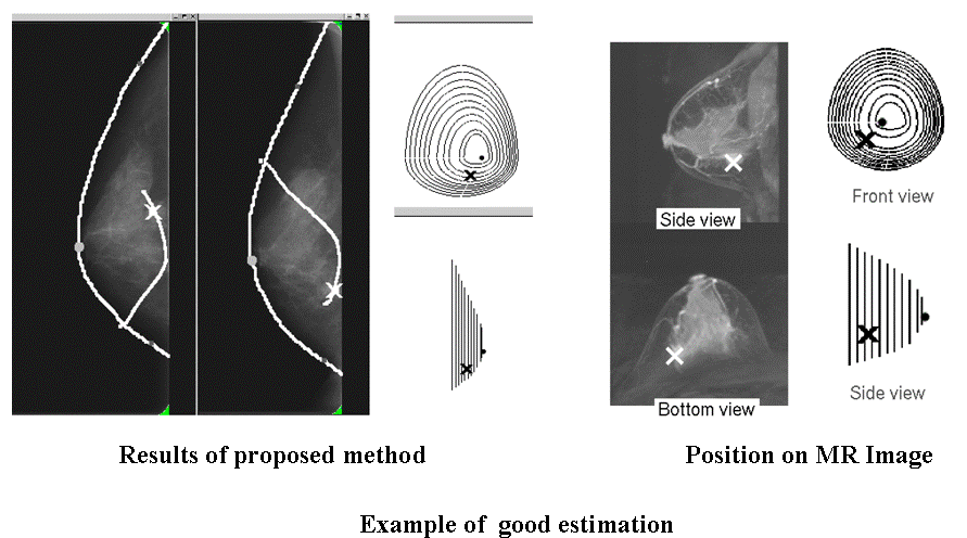
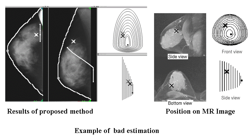
The 3D locations obtained by the system have been examined from a clinical viewpoint. We tentatively conclude that the system achieves errors within 10?20 mm in estimating the 3D locations of lesions. Most error seems to arise in the depth direction. We believe that the accuracy in the depth can be improved by replacing the simplified compression model used for the system with a richer compression model, and indeed we are continuing to work in this area.


